From time to time, we look at various types of imaging studies, such as x-rays, CAT scans, MRI, and contrast studies like angiography or barium studies that look at the GI tract. Dr. X will often ask us, "What's wrong?" As in this example:

So, this is a plain film x-ray.* Bone and metal are white, soft tissue is gray, air is black. At first glimpse, there appear to be a lot of bone fragments, little white cirles that don't seem to go with anything. In fact, this is a child. Her bones have not yet finished ossifying, so in some parts they are composed of cartlidge, which doesn't block x-rays the way the calcium in real bone does. The big thick bone coming up form the bottom of the image is the femur (thigh bone) articulating with the three bones of the pelivs. The trick with looking at pediatric images for fractures is that you have to know where it's normal to have breaks in ossification, and where it's not. One easy way is to compare the left versus right sides. A simple analysis is below:
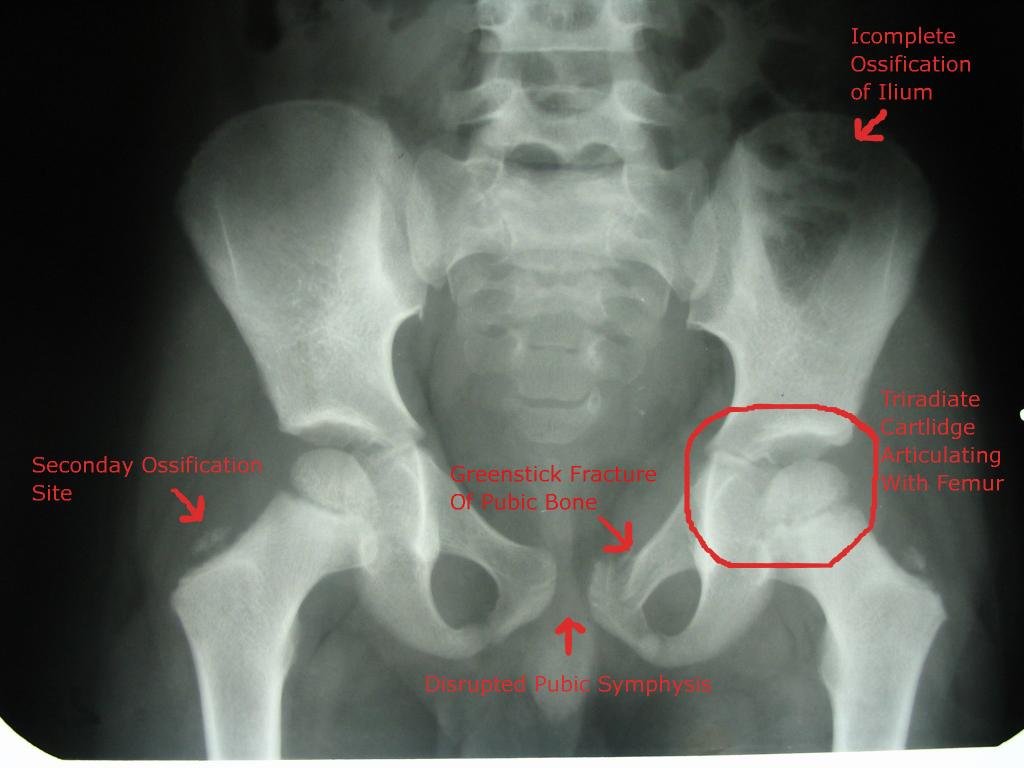
O.K., so the left pubic bone is fractured (x-rays are read as if the patient were facing you). The disrupted pubic symphysis and very slight dislocation of the left femur are results of this. Why is this my least favorite part? The answer is found in the answer to this question - what's the differential diagnosis for a pediatric fractured pubis? This is a complicated way of asking, "What could be the cause?" If you're lucky, thre's a car accident in the near past. If not, the most likely cause is child abuse.
*I don't know who this is, or the age. There are no identifying marks, therefore I consider this sanitized for the purpose of privacy. The image is a picture I took of a plain film, so it is not form any case online.

No comments:
Post a Comment